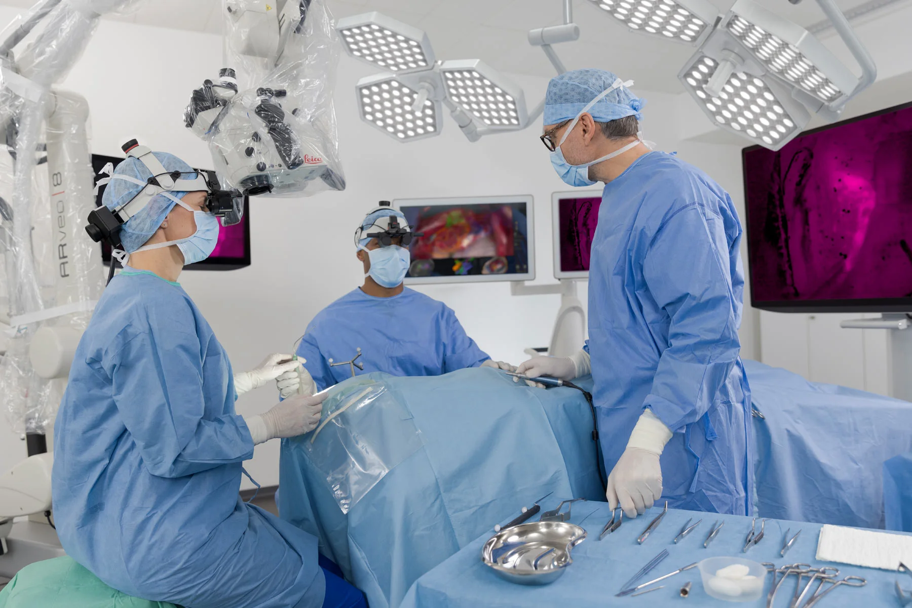
Info
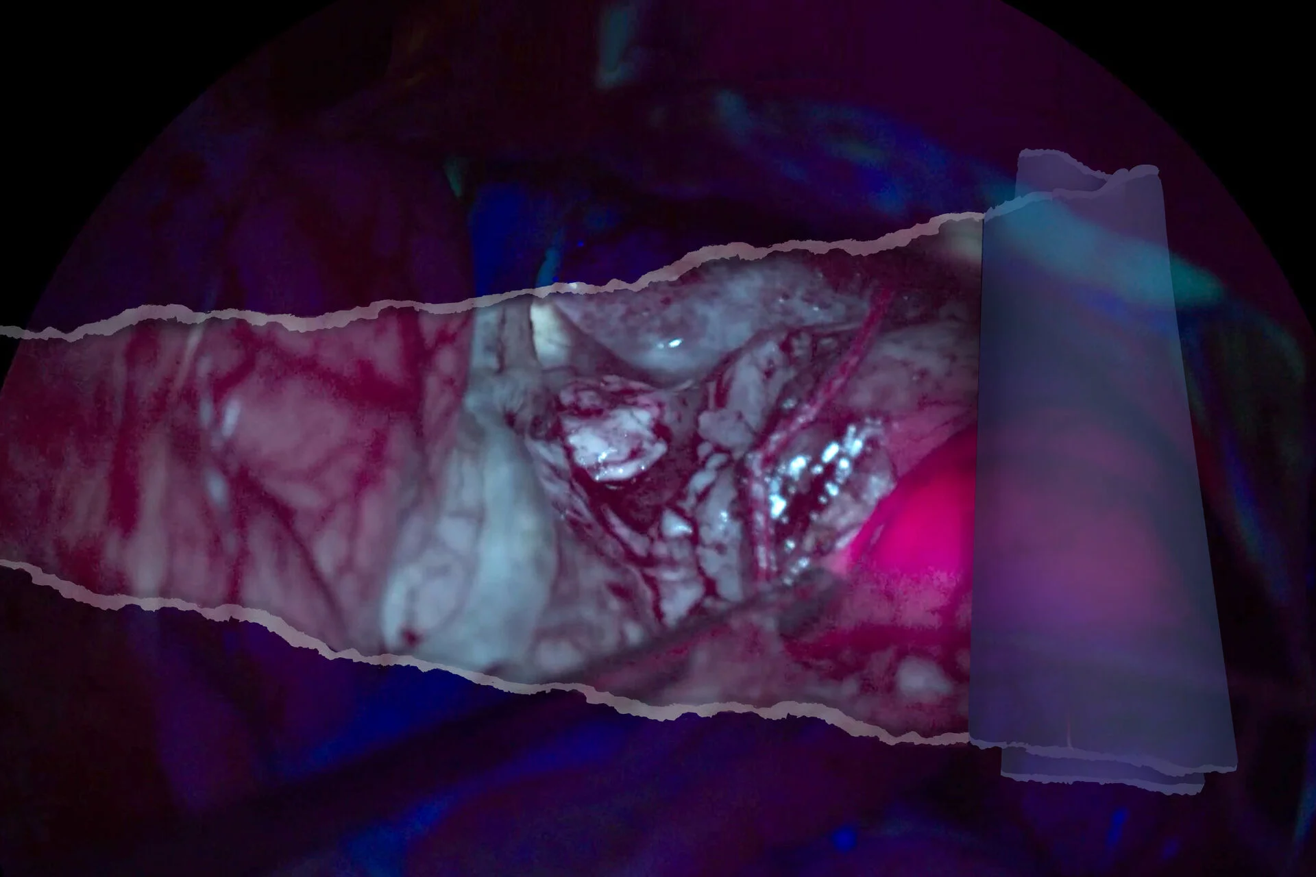
See clearer anatomical details surrounding the fluorescent-marked tumor
Enhanced visual information is essential to make precise and well-informed decisions during brain tumor surgery of suspected grade III and IV gliomas. The Anatomy view of the GLOW400 Augmented Reality fluorescence application combines the information of fluorescent and non-fluorescent tissues as well as anatomical structures—all in a single image in real time.
Advantages of the GLOW400 3D Anatomy view
- Augmenting the view of the fluorescence-marked tumor with an enhanced surrounding showing anatomical details
- Not having to recall the differences between the white light image and FL400 may reduce fatigue and minimize surgical interruptions
- Seeing more details such as vessels and bleeding that were previously hidden
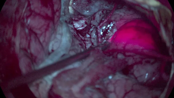
Reveal lower-intensity fluorescence signals
During tumor resection, you are dependent on being able to repeatedly check for traces of remaining visible fluorescence, particularly lower-intensity fluorescence.
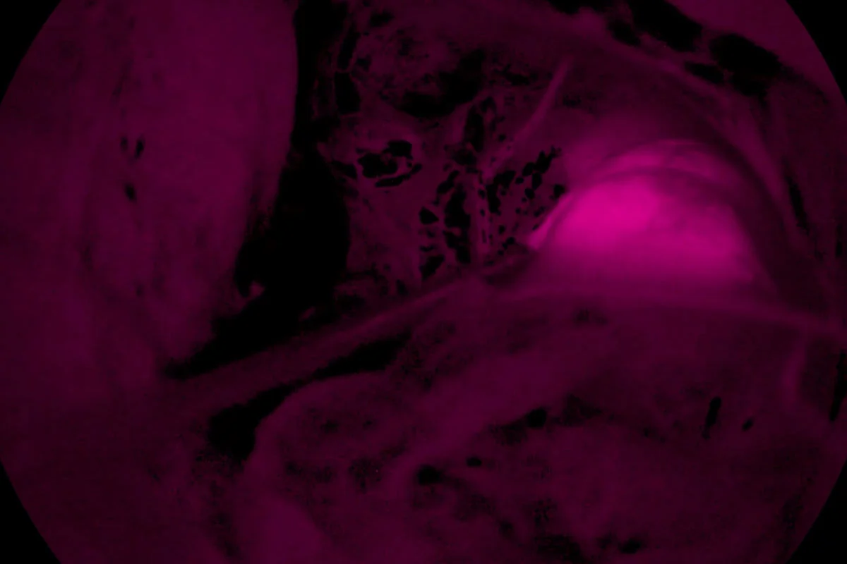
The GLOW400 Highlighted Fluorescence view transforms your visualization of suspected grade III and IV glioma tissues. Now you see what you may have previously missed.
Advantages of the GLOW400 AR fluorescence Highlighted Fluorescence view
-Broader representation of fluorescence intensities
-Enhanced visibility of low fluorescence signals to observe traces of remaining fluorescence
-Showing a pure fluorescence view on the monitor by separating fluorescent from non-fluorescent blue light signals, on a dark background
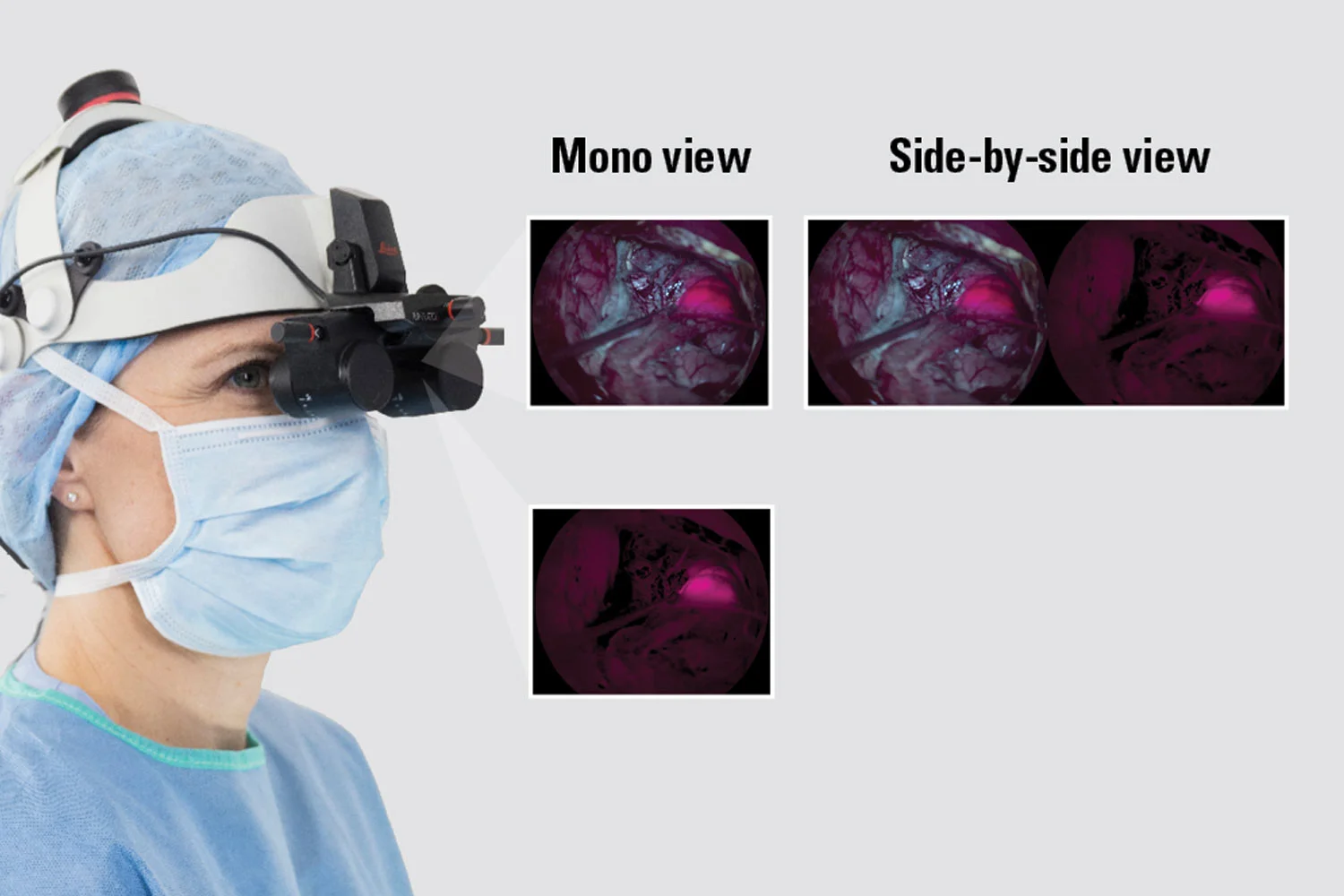
Choose how you want to view GLOW400
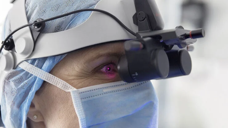
You can visualize the application on a 2D or 3D monitor and share it with your team in high resolution. You can also use the MyVeo surgical headset to view the images directly in front of your eyes, in 3D.
GLOW400 allows for seamless integration into your workflow, thanks to its compatibility with the ARveo 8 digital visualization microscope, providing you with the full benefits of integration.
*2D visualization is only for observation, not for heads-up display surgery
Choose how you want to view GLOW400
You can visualize the application on a 2D or 3D monitor and share it with your team in high resolution. You can also use the MyVeo surgical headset to view the images directly in front of your eyes, in 3D.
GLOW400 allows for seamless integration into your workflow, thanks to its compatibility with the ARveo 8 digital visualization microscope, providing you with the full benefits of integration.
*2D visualization is only for observation, not for heads-up display surgery
Toggle through multiple real-time GLOW400 views
Achieve a more comprehensive understanding of suspected grade III and grade IV glioma tissues by making comparisons of the information provided by the different views of the GLOW400.
With just a click on the microscope handle or the microscope’s footswitch, you can smoothly toggle through the mono and side-by-side views of GLOW400 Anatomy and HiFluo.
The combination of different views can reduce surgical interruptions and mental fatigue.